~ 1 min read
Chicken Breasts
In order to give a little more items of interest here, these are a few of the images which I’ve been creating to assist me with my Image Processing module here at Cardiff…
Basically, an image of a Chicken Breast is taken, and the best approximation to a cardioid found and displayed. This involves taking an image, performing Median filtering upon it to remove noise, edge detecting it with sobel masks, forming an accumulator space from the approximate gradient determined, and finally locating the largest peak within this space….
Boring eh? Take a look at a few of the images to see how it works…..I’ve impressed myself.
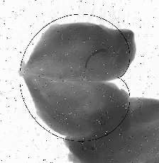
1. The original image
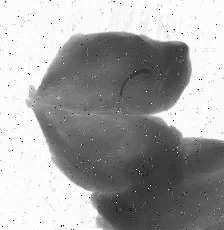
2. The median filtered image

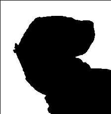
3. The thresholded image
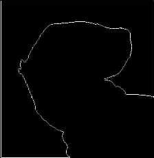
4. An edge detected version of the image
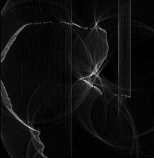
5. The accumulator image
6. The located cardioid